Fibroadenoma

March 10, 2024

Benign Breast Conditions: Meet the Fibroadenoma

Did you know that not all lumps or abnormalities found in breast tissue are cause for immediate concern? Today, we’re shedding light on a common benign (non-cancerous) breast condition known as a Fibroadenoma, often whimsically referred to as a "breast mouse."

What is a Fibroadenoma?

ο»Ώ
Fibroadenomas are solid, smooth, and firm benign tumours that are most common in women between the ages of 15 and 35. They get their nickname, "breast mouse," from their ability to move easily within the breast tissue when touched, much like a mouse scurrying under a rug.

What Causes Fibroadenomas?


The exact cause of fibroadenomas isn't fully understood, but they're closely linked to hormonal changes in the body. These benign tumours are most common during a woman's reproductive years, suggesting that estrogen plays a significant role in their development. Fibroadenomas can vary in size during the menstrual cycle, pregnancy, and breastfeeding, reflecting the influence of hormonal levels. While they are influenced by the body's hormonal environment, they are generally considered to be low risk in terms of breast cancer development.

Characteristics of Fibroadenomas:


- Size: They can range from as small as a marble to as large as a golf ball.
- Shape: Typically round or lobular, with distinct edges that sometimes can be felt through the skin.
- Feel: They are usually painless, but their presence is usually palpable and can sometimes increase in size with hormonal changes, such as during menstruation.

Why it Matters:


Understanding the nature of fibroadenomas is crucial for peace of mind and informed health decisions. While they are benign, monitoring is essential to observe any changes in size or feel, as part of routine breast health management.

Diagnosis and Management:


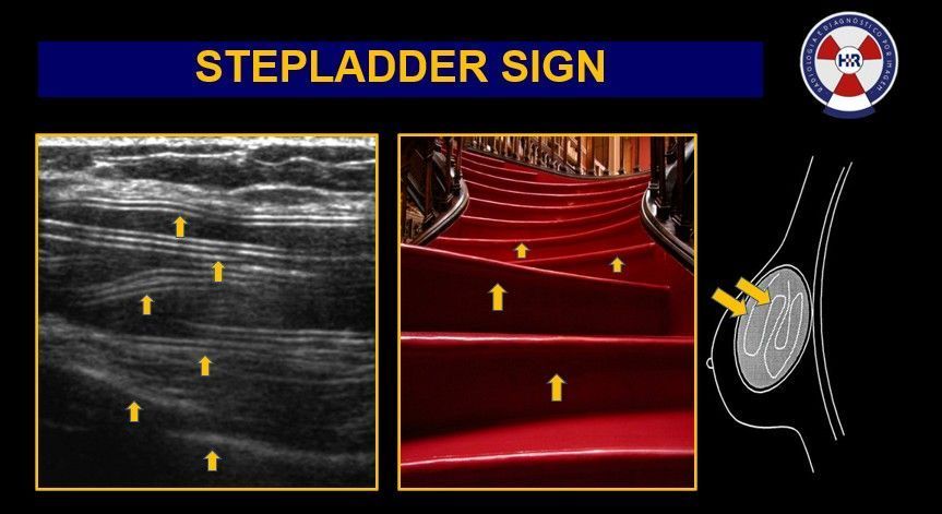
Fibroadenomas are usually diagnosed through clinical examination, ultrasound, or a mammogram and can be confirmed with a biopsy. In most cases, they don’t require treatment and may shrink on their own. It is very common to get many of them in one breast or both! However, if a fibroadenoma is large, is growing quickly or causes discomfort, it can be surgically removed.

Remember:


 Regular breast screenings and being familiar with how your breasts normally look and feel are key components of breast health. If you notice any changes or new lumps, it's important to consult with a healthcare provider.

Breast Imaging Victoria is here to support your journey towards understanding and managing your breast health. For more information or to schedule a breast scan, reach out to us today.

By Caroline O'Sullivan September 25, 2025
At Breast Imaging Victoria, we believe breast health starts with knowledge. That’s why we’re proud to offer patients access to iPrevent - an evidence-based breast cancer risk assessment tool developed by the Peter MacCallum Cancer Centre and endorsed by the The Royal Australian College of General Practitioners (RACGP) . πŸ”Ž What is iPrevent? iPrevent is a free, online tool designed to help women understand their personal breast cancer risk. It combines detailed personal and family history with validated scientific models to estimate an individual’s risk of developing breast cancer over their lifetime. ✨ Why does it matter? Not all women face the same level of risk. Age, breast density, family history, lifestyle factors, and genetic background all influence breast cancer risk. iPrevent makes it easier to translate those risk factors into meaningful, personalised information. πŸ’¬ At BIV, how do we use it? Patients have the option of completing the iPrevent risk assessment while waiting for their imaging appointment. This information helps our radiologists and multidisciplinary team personalise future imaging surveillance and make sure you receive the right level of monitoring for your risk profile. 🩺 Benefits of iPrevent at BIV: - Empowers patients with a clearer understanding of their breast cancer risk - Supports conversations with your GP or breast specialist - Guides decisions on screening frequency (annual vs biennial) and imaging type (mammogram, ultrasound, contrast-enhanced mammography) - Provides peace of mind by turning risk factors into a practical plan πŸ“£ Breast Imaging Victoria is committed to being more than just an imaging clinic — we’re a partner in your long-term breast health journey. Tools like iPrevent help us make care more proactive, personalised, and effective . You are welcome to contact us if you have further questions.
By Caroline O'Sullivan September 11, 2025
While mammograms remain a crucial part of breast screening, ultrasound is the workhorse when it comes to monitoring implant health and detecting complications. At Breast Imaging Victoria, we routinely use high-resolution ultrasound in combination with mammography to give a clearer, more complete picture. Here’s what ultrasound can help with: βœ… Silent implant ruptures It can detect small tears or leaks -even if you don’t have symptoms. For example you can get ruptures within your implant shell (intracapsular ruptures) which might not be physical noticeable but can be seen under ultrasound. A common sign is the stepladder sign as seen in the image posted. βœ… Capsular contracture Ultrasound helps measure the thickness of scar tissue forming around your implant, indicating how severe the contracture may be. βœ… Implant positioning issues We can identify signs of bottoming out, rotation, displacement or herniation. βœ… Peri-implant complications From fluid (seroma), blood (hematoma), or infection (abscess), ultrasound helps us catch issues early -before symptoms worsen. βœ… Cancer screening In augmented or dense breasts, ultrasound is often more accurate in detecting small breast masses or suspicious lesions. βœ… Avoiding unnecessary MRIs Ultrasound provides a cost-effective, radiation-free alternative for ongoing implant surveillance in many cases. βœ… Procedure guidance Ultrasound also allows precise needle guidance if a biopsy or aspiration is needed near the implant. ✨ At BIV, we tailor imaging to each patient. For women with implants and remaining breast tissue, we usually recommend both mammogram + ultrasound (unless your referral says otherwise). If you’ve had reconstruction post-mastectomy, ultrasound alone is usually appropriate. πŸ“£ Have breast implants? Talk to your GP about getting referred to a specialist breast imaging clinic. Early detection applies to everyone - and we’re here to help you stay one step ahead.
By Caroline O'Sullivan September 4, 2025
At Breast Imaging Victoria, we regularly perform specialised mammograms for women with breast implants using implant displacement views (the Eklund technique), to ensure we capture as much breast tissue as possible — especially what’s hidden behind the implant. This allows us to see up to 85% of the breast tissue, regardless of implant type or size. πŸ‘‰This specialised technique gently pushes the implant back towards the chest wall while the breast tissue is pulled forward and imaged separately. This ensures clearer, more accurate results. It may sound uncomfortable, but don’t worry — we apply soft, controlled compression to keep things as comfortable as possible while ensuring diagnostic quality. Many patients tell us they feel very little during the displaced views because the implant buffers the pressure. 🩻 Our radiographers are trained in this technique and perform it daily. These extra views are important because standard mammograms may not always see around or through the implant — especially when looking for subtle changes or early signs of cancer. πŸ‘©βš•οΈ Whether you have saline or silicone implants, both are handled the same during mammography — neither is better or worse in terms of compression or image clarity. ✨At BIV, we tailor every scan to the individual. For those with implants and breast tissue (not post-mastectomy), we usually combine mammogram and ultrasound to give the clearest picture possible. πŸ—“ The current recommendation is for women aged 45–70 to have a mammogram every 2 years - and that includes women with implants. If you have risk factors, screening may begin earlier or happen more frequently. Talk to your GP. Have questions? We’re here to help you understand your options and feel confident in your care.
More Posts